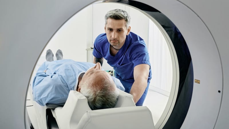
Q: I’m having an MRI on my liver and I’m frightened because I had a nasty experience the last time. There must have been something wrong with the speaker system as the radiography team couldn’t hear me, even though I was calling out because I was in pain with my arthritic shoulders (I had to hold my arms above my head). I was close to panicking with the claustrophobia. Would it be possible to have some form of sedation this time?
KS
A: Magnetic resonance imaging (MRI) is known to cause anxiety in some who find being confined inside the machine makes them feel claustrophobic.
The procedure itself is also noisy — because the scanner is essentially a giant electromagnet and makes loud clicking sounds in action — and a scan takes time.

There are steps that can help.
For shoulder pain, I recommend taking ibuprofen or paracetamol as well as codeine (available over the counter) two hours before the booked time of your scan — checking first with your GP that it’s OK for you to take those drugs.
I would also ask your GP about sedation, which is effective in calming a sense of panic. One option is a low dose of a tranquilliser (diazepam 5mg or lorazepam 1mg) — some GPs prefer to talk to the hospital and arrange for patients to receive this when they arrive for their appointment.
I would also insist on speaking to the radiographer before your scan, so you can explain the problems you had with the previous MRI.
It’s essential they don’t start scanning until you’re confident they can hear you and you can hear them when in the machine.
I don’t imagine your position as you’re scanned can be altered: the liver occupies much of the upper part of your abdomen on the right side, and lying with your arms beside you would interfere with the clarity of the images — which is why your arms need to be extended above your head.
But taking a painkiller before the scan should minimise the discomfort of undergoing this important investigation.
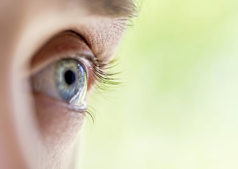
Q: I developed floaters in my left eye two years ago, then a year ago they appeared in my right eye. It’s like looking through dirty windows with dead fly splats and it’s getting me down. My specialist has said that in time they should dissipate but what are your thoughts on an operation?
YD
A: When I was a medical student back in the 1970s we called floaters muscae volitantes — the Latin for flying flies.
Essentially, they are bits of debris, formed of collagen fibres, in the vitreous humour — this is the gel that fills the eye behind the lens, giving it its shape.
These spots ‘appear’ in your vision as they move across the lens. Most of us see floaters at some point, particularly when looking at a bright surface.
The vitreous is clear when we’re born but as we age these imperfections can develop and become more intrusive, often causing cobweb-like images.
While most people are able to learn to ignore them, in others they can worsen if the vitreous humour pulls away from the retina — the light-sensitive area at the back of the eye. If this happens, it can also cause random flashes.
Your ophthalmologist will have advised you to relax and not read too much into these experiences, but I do understand how you feel about these persistent floaters. The only possible treatment is an operation, a vitrectomy.
This involves essentially suctioning out the vitreous humour and replacing it with a saline solution, which in time is replaced naturally by aqueous humour. This is the fluid that sits in front of the lens and does the job of the vitreous humour — without the floaters.
It’s unlikely you’d be offered this procedure on the NHS for floaters alone — but if the vitreous detaches from the retina, there’s a real but small risk of the retina then detaching from the back wall of the eye, something that needs prompt surgical treatment.
The symptoms of detachment are a rapid increase in floaters, flashes of light and a dark curtain forming across parts of the visual field. If you experience this, you should see a specialist quickly.
But in time, hopefully your troublesome experience with floaters will simply abate.
© Solo dmg media